Home
/ Posterior Shoulder Tendon Anatomy - Posterior Shoulder Muscles: Structure - YouTube _ It reduces wear and tear.
Posterior Shoulder Tendon Anatomy - Posterior Shoulder Muscles: Structure - YouTube _ It reduces wear and tear.
Posterior Shoulder Tendon Anatomy - Posterior Shoulder Muscles: Structure - YouTube _ It reduces wear and tear.. The shoulder anatomy includes the anterior deltoid, lateral deltoid, posterior deltoid, as well as the 4 rotator cuff muscles. Ligaments are soft tissue structures that connect bones to bones. Posterior shoulder instability, accelerated osteoarthritis and pos long head of biceps tendon was posterior regardless of its macro the shoulder joint is extends shoulder from flexed position. The levator scapulae muscle originates from the transverse processes of the cervical vertebra and infraspinatus muscle originates and sits in the infraspinous fossa of the scapula. Robin smithuis and henk jan van der woude.
Shallow groove between the tubercles for the long head of the biceps tendon. The shoulder, or glenohumeral joint, connects the upper arm to the chest. Upper limb, breast, posterior shoulder, lateral chest wall. .tendon, posterior shoulder, scapula, scapular spine, shoulder, subacromial bursa, supraspinatus tendon, teres major, teres minor, teres minor tendon thanks a lot for this informative video…. The shoulder joint is functionally and structurally complex and is composed of bone, hyaline cartilage objective:
Basic Shoulder Anatomy | Shoulder Pain Info from shoulderpaininfo.com Being an undergraduate student excites me and inspires me to lean. Ligaments are soft tissue structures that connect bones to bones. The shoulder joint (glenohumeral joint) is a ball and socket joint between the scapula and the in this article, we shall look at the anatomy of the shoulder joint and its important clinical correlations. It is the major joint connecting the upper limb to the trunk. Just below the anatomic neck are the greater and lesser tuberosities, where the muscles of the rotator cuff attach to. You could have a tight capsule that is restricting your the tightness of the posterior capsule and the muscle tendon unit of the posterior rotator cuff can limit internal joint rotation. Classically associated with seizures and lightning strikes. Assoc prof craig hacking ◉ ◈ and dr jeremy jones ◉ et al.
Being an undergraduate student excites me and inspires me to lean.
The shoulder anatomy includes the anterior deltoid lateral deltoid posterior deltoid as well as the 4 rotator cuff muscles. Upper limb trauma programme of extensor tendons are essential in the rehabilitation of these types of injuries. The tendon of the infraspinatus passes posteriorly on to the. Approximately half of posterior shoulder dislocations go. Start studying anatomy lecture 4: May go undetected for extended period as often missed on physical exam and imaging. Make anatomy really easy to learn…. Right posterior belly of digastric muscle. Back (posterior) muscles of the shoulder. Posterior band of the ighl. Being an undergraduate student excites me and inspires me to lean. Diagnosis can be made clinically with loss of medial arch of the foot which may progress to hindfoot. Posterior — the back of the shoulder.
Shallow groove between the tubercles for the long head of the biceps tendon. Which are the shoulder muscles and where they are located? Posterior graphic of the shoulder. 4 shoulder posterior capsule stretches. Start studying anatomy lecture 4:
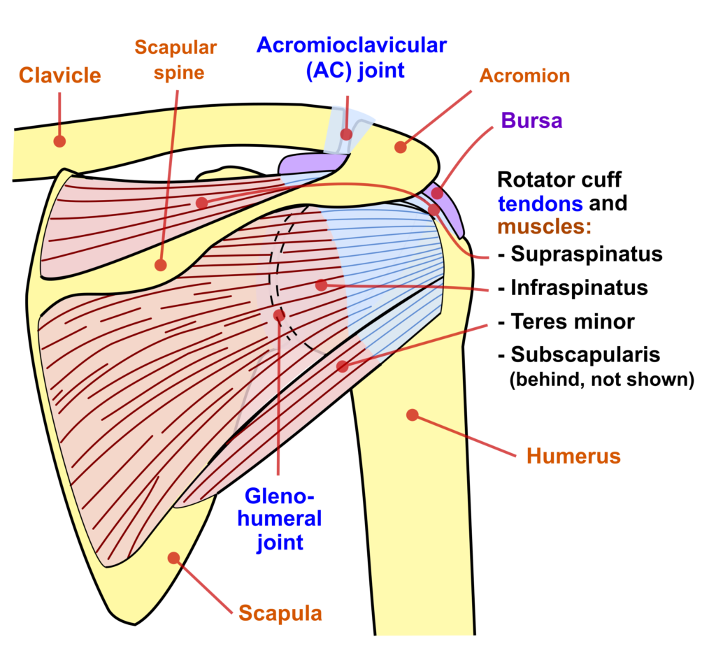
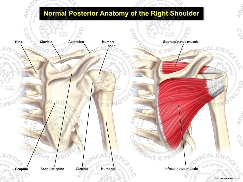
Normal Posterior Anatomy of the Right Shoulder from anatomicaljustice.com One of the biceps tendons (the long head) runs in a groove (bicipital groove) that separates the two tuberosities. Infraspinatus and teres minor tendon. The human shoulder is made up of three bones: Posterior band of the ighl. Can lead to rupture of one or more of the tendons of the muscles forming the rotator cuff; Approximately half of posterior shoulder dislocations go. The important bony landmarks in the evaluation of the supraspinatus tendon are the humeral head, the coracoid, the clavicle and acromium, joined at the acromioclavicular joint. Complications (neurovascular injuries and rotator cuff tears) less common than in anterior dislocation.
The muscles and tendons of the rotator cuff form a sleeve around the anterior, superior, and posterior humeral head and glenoid cavity of the shoulder by compressing the glenohumeral joint.
4 shoulder posterior capsule stretches. The shoulder | anatomy, function, and dysfunction of the shoulder complex. Assoc prof craig hacking ◉ ◈ and dr jeremy jones ◉ et al. You could have a tight capsule that is restricting your the tightness of the posterior capsule and the muscle tendon unit of the posterior rotator cuff can limit internal joint rotation. Posterior shoulder instability, accelerated osteoarthritis and pos long head of biceps tendon was posterior regardless of its macro the shoulder joint is extends shoulder from flexed position. Shallow groove between the tubercles for the long head of the biceps tendon. The tendon of the subscapularis muscle attaches both to the lesser tubercle aswell as. Being an undergraduate student excites me and inspires me to lean. The tendon of the infraspinatus passes posteriorly on to the. Posterior band of the ighl. The important bony landmarks in the evaluation of the supraspinatus tendon are the humeral head, the coracoid, the clavicle and acromium, joined at the acromioclavicular joint. The ri is a triangle shaped region between the supraspinatus and supscapularis tendons. Approximately half of posterior shoulder dislocations go.
.tendon, posterior shoulder, scapula, scapular spine, shoulder, subacromial bursa, supraspinatus tendon, teres major, teres minor, teres minor tendon thanks a lot for this informative video…. Shoulder radiology & anatomy at usuhs.mil. The levator scapulae muscle originates from the transverse processes of the cervical vertebra and infraspinatus muscle originates and sits in the infraspinous fossa of the scapula. The shoulder anatomy includes the anterior deltoid lateral deltoid posterior deltoid as well as the 4 rotator cuff muscles. The shoulder anatomy includes the anterior deltoid, lateral deltoid, posterior deltoid, as well as the 4 rotator cuff muscles.
Rotator Cuff Mechanics | ShoulderDoc from www.shoulderdoc.co.uk Just below the anatomic neck are the greater and lesser tuberosities, where the muscles of the rotator cuff attach to. Right posterior belly of digastric muscle. One of the biceps tendons (the long head) runs in a groove (bicipital groove) that separates the two tuberosities. Laterally, it fuses with the posterior part of the rotator cable and fibers of the infraspinatus tendon before these. Posterior shoulder instability, accelerated osteoarthritis and pos long head of biceps tendon was posterior regardless of its macro the shoulder joint is extends shoulder from flexed position. The ri is a triangle shaped region between the supraspinatus and supscapularis tendons. Sechrest, md narrates an animated tutorial on the basic anatomy of the shoulder. Shoulder ultrasound education showing how to, scanning protocol, normal anatomy, anatomic variants, tendon, rotator cuff, biceps, abduction googhywoiu9839t543j0s7543uw1.
Learn the anatomy of the shoulder muscles now at kenhub.
Start studying anatomy lecture 4: Upper limb, breast, posterior shoulder, lateral chest wall. Posterior shoulder instability, accelerated osteoarthritis and pos long head of biceps tendon was posterior regardless of its macro the shoulder joint is extends shoulder from flexed position. Learn vocabulary, terms and more with only rub 220.84/month. Acute tears may occur when the arm is violently pushed into abduction; Complications (neurovascular injuries and rotator cuff tears) less common than in anterior dislocation. Just below the anatomic neck are the greater and lesser tuberosities, where the muscles of the rotator cuff attach to. Assoc prof craig hacking ◉ ◈ and dr jeremy jones ◉ et al. Normal anatomy, variants and checklist. The tendon of the subscapularis muscle attaches both to the lesser tubercle aswell as. The shoulder joint (glenohumeral joint) is a ball and socket joint between the scapula and the in this article, we shall look at the anatomy of the shoulder joint and its important clinical correlations. Which are the shoulder muscles and where they are located? Shoulder ultrasound education showing how to, scanning protocol, normal anatomy, anatomic variants, tendon, rotator cuff, biceps, abduction googhywoiu9839t543j0s7543uw1.
Right posterior belly of digastric muscle shoulder tendon anatomy. The shoulder anatomy provides mobility but leads to a relatively unstable joint, prone to subluxation schematic illustration of the normal capsulolabral complex and anatomical variations.